
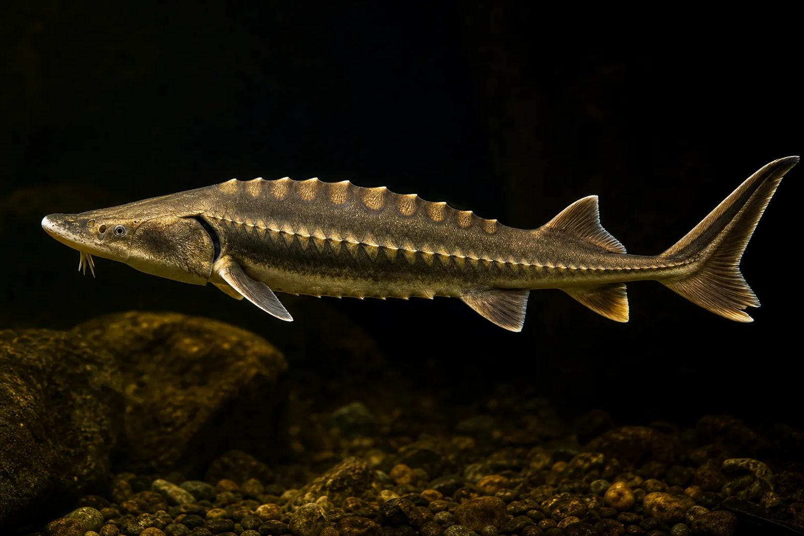
Five rows of bony armour run along a sturgeon's body. Beneath its snout, four fleshy barbels help locate food; a toothless mouth extends to suck it in. Inside, a largely cartilaginous skeleton supports the fish. The organs that matter most for caviar lie deeper still: paired ovaries, where eggs grow attached to supporting tissue.
Sturgeon anatomy at a glance
| Feature | What it does | Why it matters for caviar |
|---|---|---|
| Rostrum, barbels and electroreceptors | Help locate food | Explain feeding, not egg quality. |
| Scutes | Protect the body | Cannot reveal ovarian maturity. |
| Tail and fins | Propel, steer and stabilise | Describe the living fish, not a caviar grade. |
| Notochord and cartilage | Support the body | Distinct from the bony skin plates. |
| Gills | Extract oxygen from water | Handle respiration, unlike the swim bladder. |
| Swim bladder and spiral intestine | Aid buoyancy and digestion respectively | Neither produces eggs. |
| Ovaries and testes | Produce eggs and sperm respectively | Examination can help distinguish females from males. |
Snout, sensors and mouth
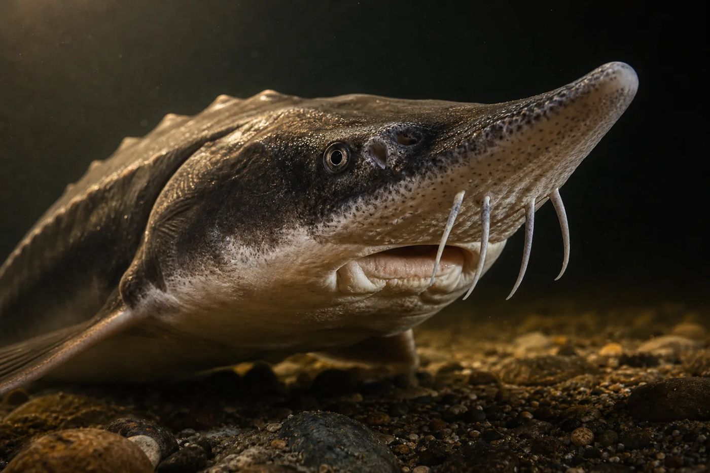
A sturgeon's snout carries an array of food-finding sensors. The rostrum projects in front of the eyes, with four barbels hanging underneath, ahead of the mouth. These fleshy sensory appendages help locate food. They neither serve as teeth nor grasp prey like fingers.
Electroreceptors give the fish another source of information. Tiny sensory structures called ampullary organs detect weak electric fields in the water, including those associated with other animals. They form part of the lateral line sensory system alongside neuromasts, which detect local water movement. Research on developing sterlet has mapped both types of organ using gene expression markers, tracing two distinct senses: electrical detection and movement detection.
The adult mouth is toothless and protrusible: it can extend outwards. Set beneath the head, it allows bottom-feeding species to bring the opening towards food and draw it in by suction. NOAA's account of Atlantic and shortnose sturgeon describes them swallowing prey whole and expelling unwanted sand and gravel.
Skin armour, fins and tail
A sturgeon's ridges are armour built into its skin. Called scutes, these enlarged bony plates are modified scales, quite unlike the thin, overlapping scales of many other fish. Their characteristic layout is five longitudinal rows: one along the back, one along each side and two towards the underside.
The scutes protect the fish, and their prominence can change with age. FAO's Siberian sturgeon account describes sharply tipped scutes in young fish but not adults. This external bone sits alongside a mostly cartilaginous internal skeleton: different tissues, in different parts of the body.
How scutes attach to the skin also matters to the disputed kosher question. The Orthodox Union rejects sturgeon, while some Conservative Jewish authorities permit it.
The tail supplies propulsion. Its caudal fin is heterocercal, with an upper lobe larger than the lower and the body's supporting axis extending into it. Paired pectoral fins behind the head and pelvic fins farther back help with manoeuvring and balance. The single dorsal fin above and anal fin below help keep the fish stable.
Inside the body
Cartilage forms much of a sturgeon's internal framework, but that does not make it a shark. Bone is present in parts of the head and shoulder region as well as in the scutes. Along the body, beneath the spinal cord, runs the persistent notochord: a flexible supporting rod.
An ovary is therefore more than a bag of loose, finished caviar.
That rod remains a major part of the adult sturgeon's axial support, unlike the chain of fully formed bony vertebral bodies familiar from many fishes. Notochord and spinal cord have separate roles: the former is supporting tissue, the latter nervous tissue. The broader evolutionary story belongs in the living fossil account.
Just behind the head, an enlarged bony flap called the subopercle covers the gills; the usual opercle bone is absent. Water passing over the gills supplies oxygen. Buoyancy is the job of a different organ, the swim bladder, a gas-containing sac. In sturgeons this bladder is physostomous, meaning it retains a connection to the digestive tract.
Following that tract farther into the body brings us to the intestine and its spiral valve. This internal fold increases the surface available for absorption and slows the passage of food. Its work is digestion, separate from the reproductive organs that share the body cavity. Eggs do not travel through it.
Where the eggs develop
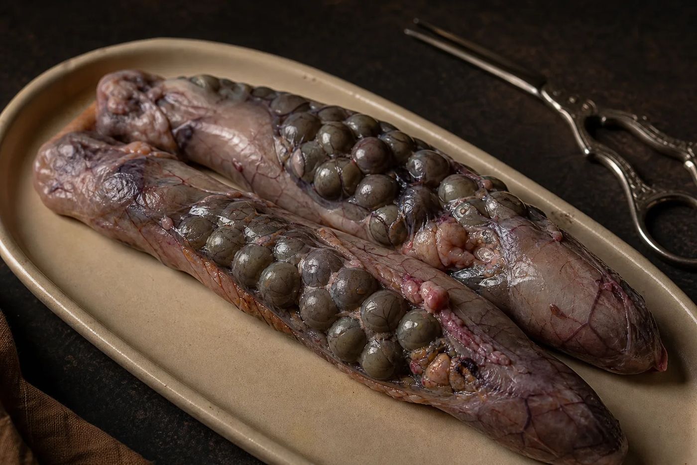
The eggs destined for caviar develop in paired ovaries inside the trunk. Ovaries in females and testes in males are the gonads, the reproductive organs. Both are elongated structures lying along the sides of the body cavity, attached to its dorsal wall. A farm worker examining them is looking for organs inside the fish's trunk, rather than an external pouch of eggs.
Within the ovaries, folds of tissue bear developing oocytes, or egg cells. Connective tissue holds the eggs together in what processors call the skein. Within this assembly, each developing egg remains associated with its own follicle, a layer of supporting cells. As development progresses, the individual eggs become increasingly conspicuous. The ovaries enlarge too, eventually occupying much of the body cavity in a mature female.
Ovulation changes that attachment: the egg is released from its follicle. This is the anatomical distinction between collecting ovarian eggs, still associated with supporting tissue, and collecting eggs after ovulation. An ovary is therefore more than a bag of loose, finished caviar. Egg development and atresia explores those stages and the reabsorption of eggs.
Mature testicular tissue looks quite different: smooth and pale, in contrast to the visible beads of developing eggs. Testes enlarge as sperm production progresses, and their appearance changes with reproductive stage. They produce sperm, carried in the reproductive fluid called milt.
How anatomy guides caviar production
To assess sex and reproductive development, farmers look inside the fish. Its outer shape cannot provide the same information as examining the gonads. Ultrasound reveals internal structures; endoscopy allows direct inspection through a small opening; biopsy removes a tissue sample. Each method answers related but different questions.
Ultrasound images depend on how sound travels through muscle, fat and gonadal tissue. FAO's hatchery manual illustrates the changing appearance of ovaries and testes across maturity stages. It also shows why the tissue surrounding a gonad can matter when interpreting an image: a visible mass is not necessarily tissue that produces eggs.
Egg collection follows the anatomy. Conventional processing separates ovarian eggs from the skein, while collection after ovulation begins with eggs already released from their follicles. For examination techniques, their limitations and required access, see sexing sturgeon; the anatomy here provides background, not instructions for handling a live fish.
What anatomy can tell you about species
Identifying a sturgeon means reading several features together. A snout may be long, short, pointed or broad. Barbels may be smooth or fringed, and their position between the mouth and snout tip can be revealing. The number and arrangement of scutes, along with mouth shape and lip structure, also contribute. A silhouette alone is not enough.
Fisheries and Oceans Canada's identification guide offers a useful comparison. White sturgeon have barbels closer to the snout tip; green sturgeon have them midway between mouth and tip, or nearer the mouth. The guide's illustrations combine this difference with scute arrangement and features on the underside, showing how several characters work together.
Some fish remain difficult to identify by appearance. An expert identification review submitted to CITES documents overlap in countable features, changing proportions during growth and particular difficulties with hybrids. A rounded snout or a scute count can support an identification without independently establishing every fish's ancestry. Where morphology is inconclusive, genetic analysis may be needed.
For the species accounts and their distinguishing features, start with sturgeon biology. Consider the fish as a whole when comparing individual features. Once the head, skin and fins are absent, the external characters described here are no longer available to examine.
Sources
-
FAO. Chebanov, M. S. and Galich, E. V. Sturgeon Hatchery Manual. FAO Fisheries and Aquaculture Technical Paper 558, 2013 edition. General anatomy and the illustrated atlas of reproductive development and ultrasound examination. Retrieved PDF.
-
Florida Museum of Natural History, University of Florida. Acipenseridae (Sturgeons). Undated web edition, accessed November 2026. Family-level anatomical characters, including scutes, barbels, mouth and adult dentition. Retrieved page.
-
NOAA Fisheries. SCUTES Student Resources. Undated web edition, accessed November 2026. Sections on feeding, fins, scutes and gills. Retrieved page.
-
Minařík, M. and colleagues. Identification of multiple transcription factor genes potentially involved in the development of electrosensory versus mechanosensory lateral line organs. Frontiers in Cell and Developmental Biology, volume 12, article 1327924, 2024. Published version, 18 March 2024. Retrieved article PDF.
-
FAO. Acipenser baerii. Cultured Aquatic Species Fact Sheets, 2009 edition. “Biological features” section. Retrieved page.
-
University of Florida, IFAS Extension. Technically Speaking, What Is Sturgeon Caviar? Publication FA194. Web edition retrieved November 2026; revision year not established from the accessible record. Sections on the female ovary, follicles and egg maturation. Retrieved indexed page.
-
University of Florida, IFAS Extension. Sturgeon Aquaculture: Specialized Techniques: Determining the Sex of Sturgeon by Direct Examination of the Gonad Using a Minimally Invasive Surgical Procedure. Publication FA183. Web edition retrieved November 2026; revision year not established from the accessible record. Gonadal appearance and direct examination. Retrieved page.
-
Fisheries and Oceans Canada. Sturgeon identification guide: Sturgeon found in British Columbia waters. Web edition, accessed November 2026. Barbel position, scutes and ventral identifying features. Retrieved page.
-
CITES Secretariat. SC74 Doc. 47, identification review submitted to the 74th Standing Committee meeting, 2022. Sections on morphological identification, age limitations and hybrids. Retrieved document.
-
Billard, R. and Lecointre, G. Biology and conservation of sturgeon and paddlefish. Reviews in Fish Biology and Fisheries, volume 10, 2001. Published review, general morphology section. Retrieved PDF.
-
Scott, W. B. and Crossman, E. J., Fisheries Research Board of Canada. Freshwater Fishes of Canada. Bulletin 184, 1973. Sturgeon anatomical descriptions, including persistent notochord and absence of vertebral centra. Retrieved government-hosted extract.
-
Orthodox Union. Goldberg, C. Kosher Fish Production: Getting Into the Stream of Things. Published 24 January 2022. The Orthodox Union's position on sturgeon scutes and their attachment to the skin. Retrieved page. My Jewish Learning. Ask the Expert: Fins and Scales. Undated web edition, retrieved November 2026. Reports the differing Conservative position. Retrieved indexed page.
-
The New International Encyclopædia. Skull of a sturgeon with membrane bones removed. Volume 18, 1905, page 642; artist unidentified. Digital reproduction and rights record held on Wikimedia Commons. Retrieved image record.

This piece was researched using anatomical literature, public-agency identification resources, university aquaculture guidance and a rights-checked historical illustration.
Last checked: November 2026